
The equipment behind an accurate diagnosis.
Every treatment decision at Aadithya Eye Care begins with a thorough examination. Our diagnostic tools let us see what the eye alone cannot reveal, so problems are caught early and treatment is planned with confidence.
- 6+
- Diagnostic and surgical tools
- 25+
- Years using clinical equipment
- All ages
- Child to senior, every visit
Good treatment starts with a clear picture.
The right diagnosis is not just the first step in eye care, it is the most important one. Equipment that shows the eye in accurate detail gives us the information to make confident decisions and to catch conditions before they become harder to treat.
At Aadithya Eye Care, every consultation is supported by clinical instruments that have been proven over years of practice. We do not guess when we can measure, and we do not treat what we have not seen clearly first.
- Catches changes before symptoms appear
- Supports honest, evidence-based advice
- Tracks progress over years of follow-up
- All in-house, at a single visit
Tools that let us see exactly what is happening.
Each instrument has a specific job. Together they give us a complete clinical picture of your eyes, from the outermost surface to the deepest layers of the retina.
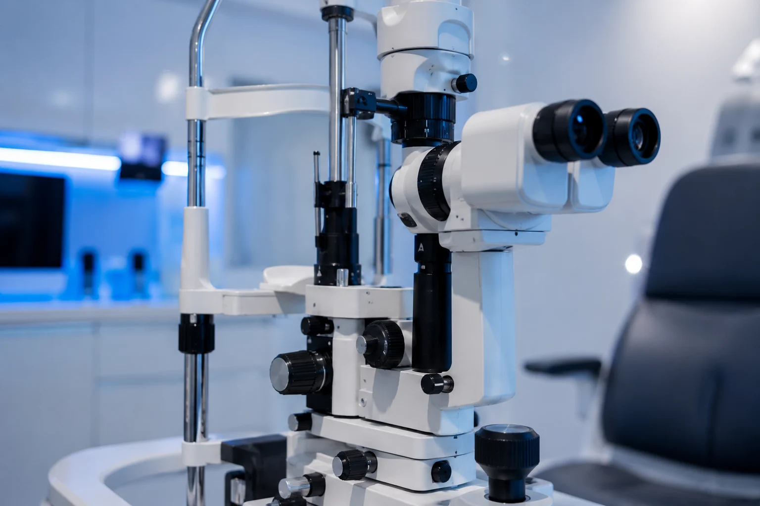
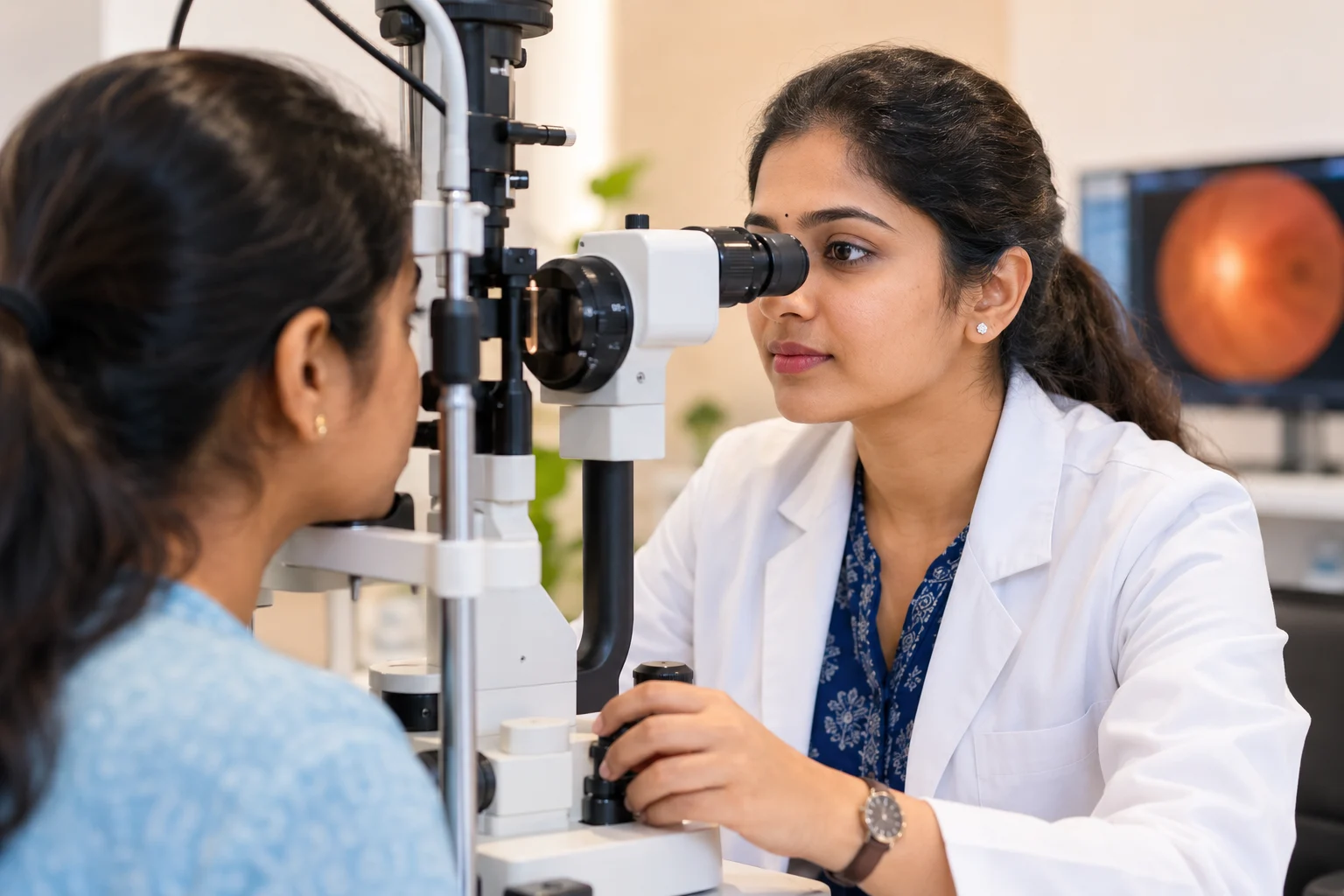
Slit-Lamp Biomicroscopy
The slit lamp is the centrepiece of every eye consultation. A focused beam of light and high magnification give us a detailed, three-dimensional view of the eyelids, cornea, lens, iris and anterior chamber.
It is how we spot corneal infections, early cataracts, lens changes and anterior segment disease that would otherwise be invisible to the naked eye.
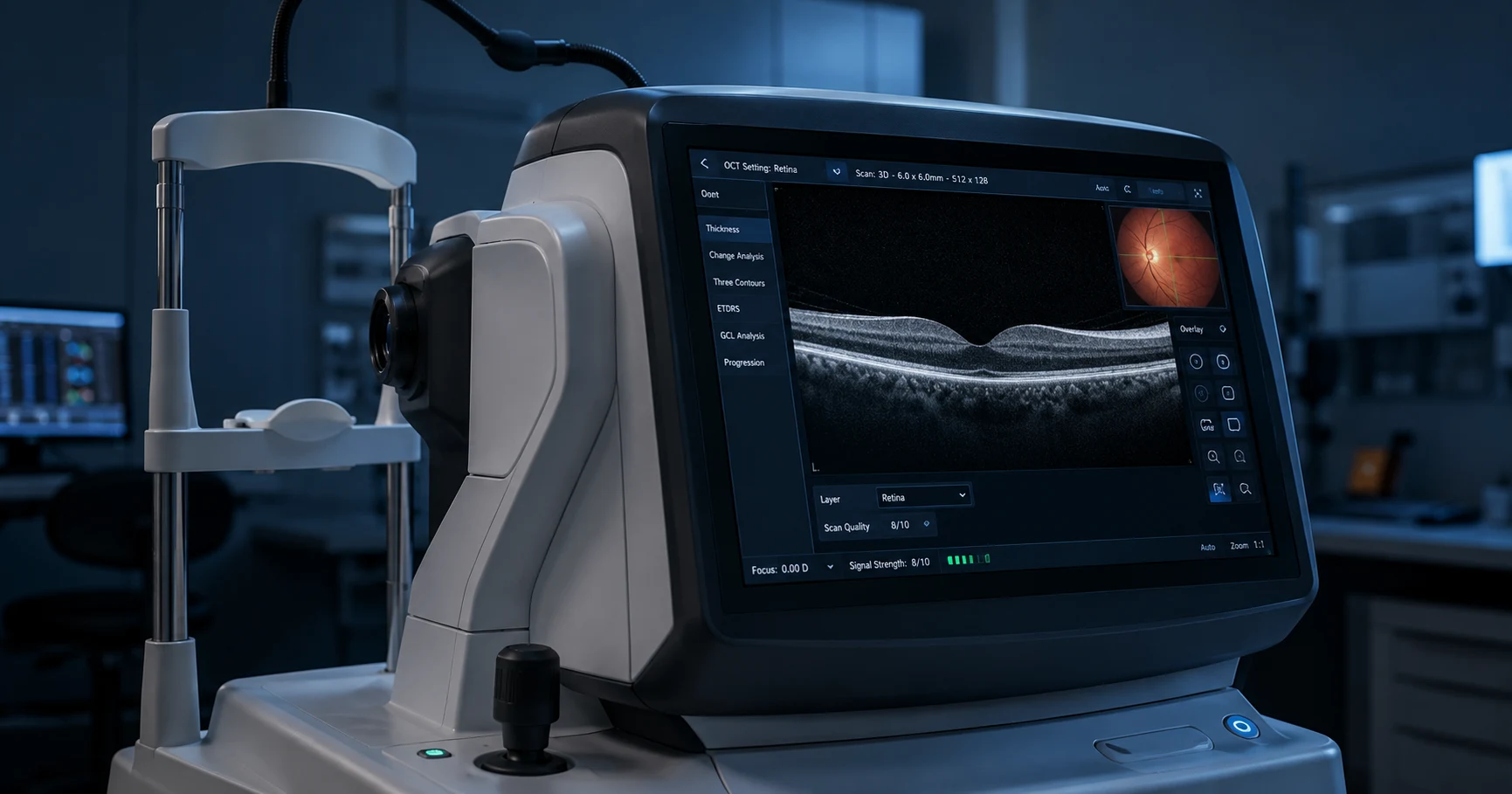
OCT Retinal Imaging
Optical coherence tomography produces cross-sectional scans of the retina and optic nerve with microscopic resolution. It lets us see individual retinal layers that are invisible in a standard fundus examination.
OCT is how we detect early macular degeneration, glaucomatous optic nerve damage and diabetic retinal swelling before they affect your central vision.
Goldmann Tonometry
Intraocular pressure is the primary risk factor for glaucoma. Goldmann applanation tonometry is the gold standard for measuring it accurately, far more reliable than the air-puff methods used at optical shops.
Every patient with a risk factor for glaucoma has their pressure checked at each visit so we can track trends, not just individual readings.
Perimetry
Perimetry maps the full visual field to detect and quantify blind spots. Glaucoma characteristically damages peripheral vision first, and perimetry is the way we find and measure that damage before it reaches the centre.
Results are compared across visits to determine whether a condition is stable or progressing, which directly guides treatment decisions.
Fundus Examination and Photography
A dilated fundus examination gives us a wide view of the retina, optic disc and blood vessels at the back of the eye. Fundus photography records what we see so findings can be compared across months and years.
This is the primary tool for monitoring diabetic retinopathy, hypertensive changes, retinal tears and disc changes in patients we follow long term.
Biometry and Keratometry
Before cataract surgery, precise measurements of the eye's axial length and corneal curvature are essential to calculate the correct intraocular lens power.
Accurate biometry is what determines how close to spectacle-free a patient's vision will be after surgery. We take these measurements carefully and confirm them before any lens implant decision is made.
What happens during a full eye assessment.
A complete eye examination at Aadithya Eye Care takes time and follows a logical sequence. Here is how it typically unfolds so you know what to expect before you arrive.
Vision assessment and refraction
We start with your best-corrected visual acuity and, where relevant, a full refraction to check whether your current glasses or contact lens prescription needs updating.
Eye pressure measurement
Intraocular pressure is measured with Goldmann tonometry at every full assessment, not only when glaucoma is suspected.
Slit-lamp examination of the front of the eye
The lids, cornea, lens and anterior segment are examined in detail under magnification. This is where corneal disease, early cataracts and lid conditions are identified.
Dilated fundus examination
Where clinically indicated, drops are used to widen the pupil so the retina, optic disc and blood vessels can be examined properly. You will need someone to help you home if you are dilated.
Findings explained and next steps discussed
Before you leave, everything we found is explained clearly. Any imaging is shown to you. If further tests or treatment are needed, we discuss the options without pressure.
nothing skipped
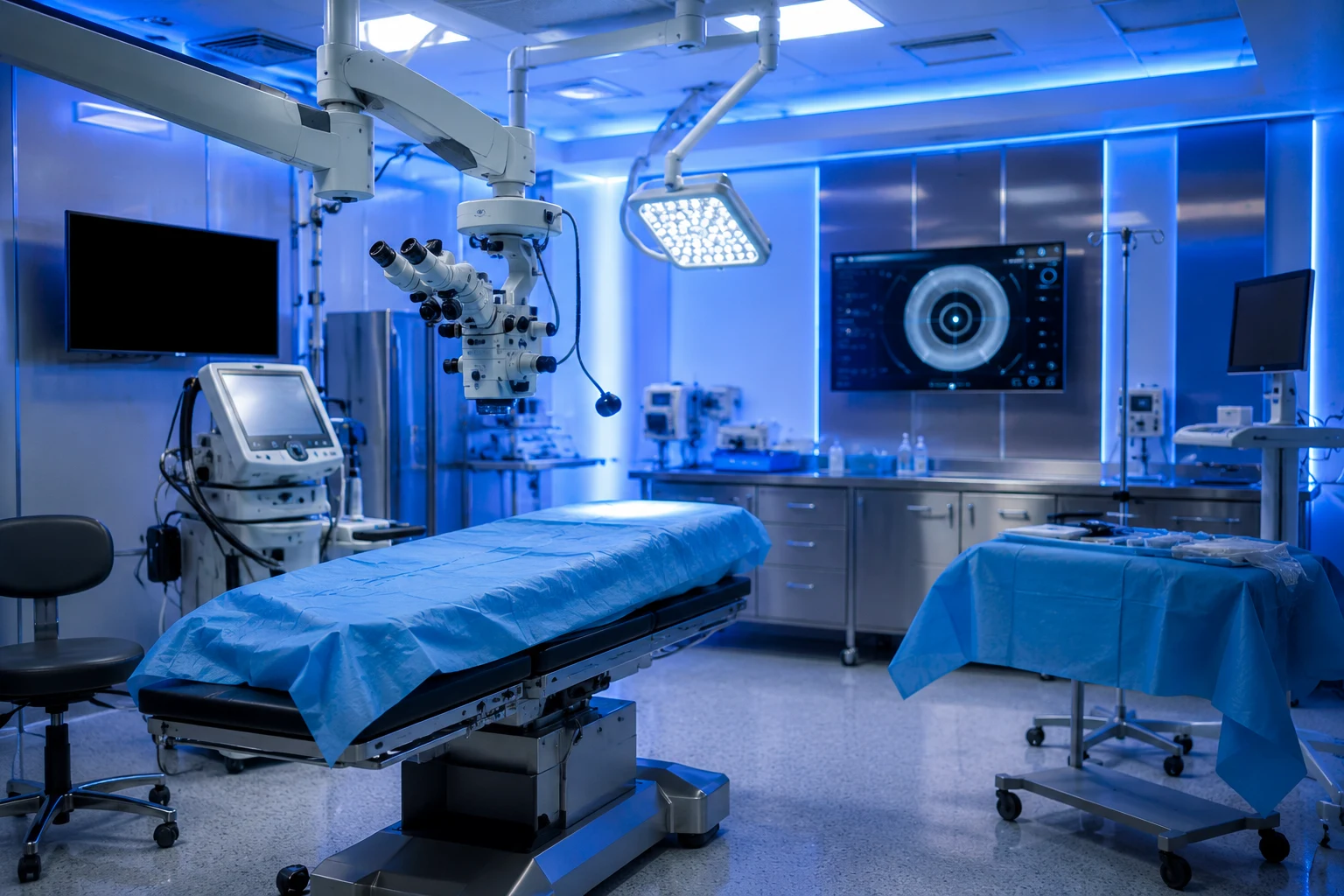
Surgery, arranged at a partner hospital.
Eye surgery needs a controlled, sterile environment and full continuity of care. We do not have an operating theatre on site, so when surgery is needed, we arrange it at a partner hospital, and our team stays with you throughout your pre-operative assessment, the surgery itself and your recovery. The phacoemulsification technique allows stitch-free cataract removal through a microincision, with most patients able to resume normal activities within days.
Accredited facilities
Surgery arranged for you with complete sterility protocols and infection control.
Modern techniques
Stitch-free cataract surgery using phacoemulsification with micro-incisions.
Continuous monitoring
Vital signs tracked and continuous care throughout the procedure.
Dedicated surgical team
Experienced ophthalmologists and clinical staff present for every case.
Continuity of care
From pre-op assessment through post-operative recovery, our team guides you every step.
See what is happening inside your eyes.
A thorough eye examination takes less than an hour and can catch conditions that have no early symptoms. Our team will call you to confirm a time that suits you.